2000B: Anasomes
|
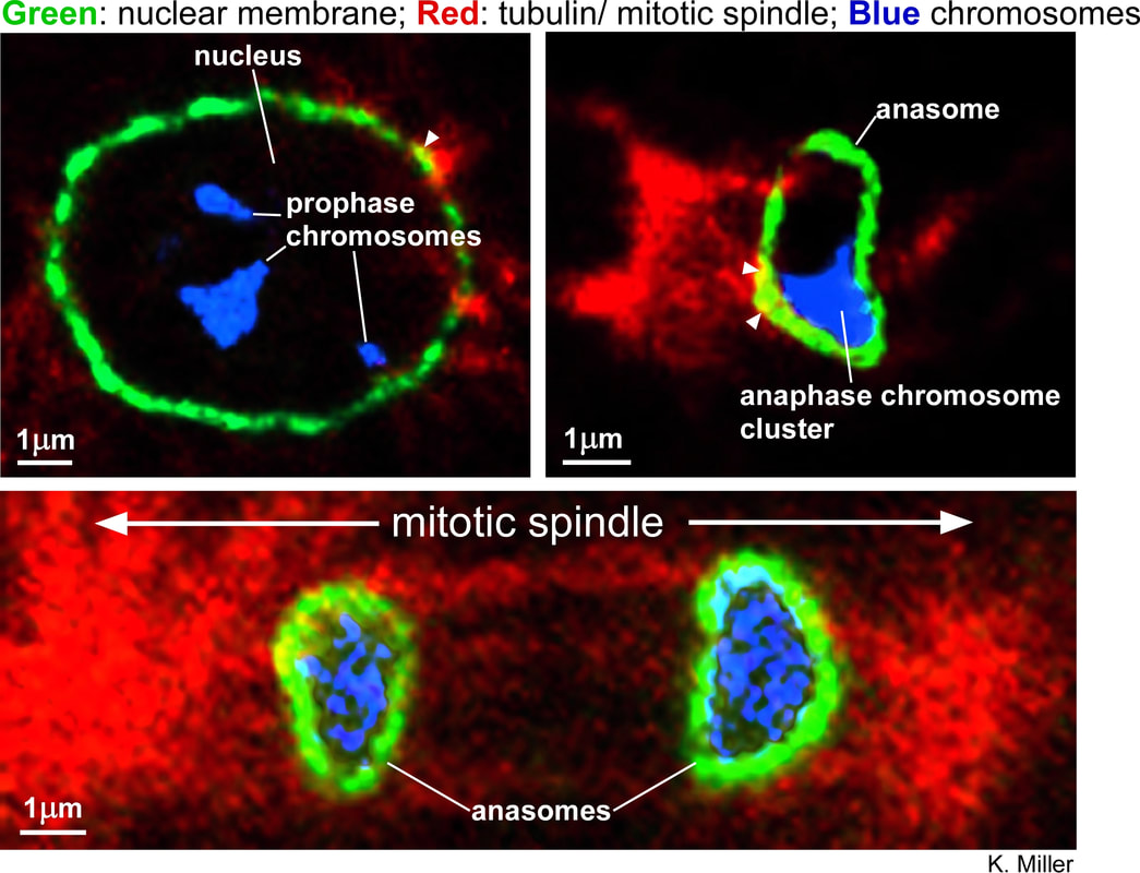
While attempting to immunostain for RIC-8 in young embryos, we found that our antibody appear to cross react with the nuclear membrane. We think this is cross-reactivity rather than bonified RIC-8 staining because the staining appeared relatively unaffected by a ric-8 mutation that strongly decreased neuronal staining. Since the cells of the early embryo divide every 15 min or so, it was fairly common to catch cells in the formaldehyde-fixed embryos in various stages of mitosis. We triple stained with the nuclear membrane antibody in green, DAPI (for DNA/ chromosomes) in blue, and tubulin (for centrosomes and microtubule asters) in red. Normally, the nuclear membrane is supposed to break-down in late prophase (the first stage of mitosis) and not re-form until telophase (the last stage of mitosis when the chromosomes are completely pulled apart and the mitotic spindle disassembles. Unexpectedly, we observed an intact nuclear membrane around the chromosomes as they were being pulled apart during anaphase, the stage of mitosis that precedes telophase and before the mitotic spindle had broken down. Upon close inspection, the anaphase nuclear membrane staining appeared to completely encircle the chromosome set. Regions of overlap with tubulin staining (which show up as yellow) suggest that the microtubules make contact with the cytoplasmic side of the nuclear membrane. Microtubules do not appear to penetrate the structure. It appears as if the microtubules, rather than being attached to the kinetochores of the individual chromosomes, are instead attached to and pulling on the nuclear membrane surrounding the tightly clustered chromsosomes. Since this anaphase structure has not, to our knowledge, previously been described, we refer to it as the "anasome". |

|
It seems that the spindle microtubules would have to initially attach to the kinetochores on the individual chromosomes in order to pull the sister chromosomes apart. If our observations are correct, and there is indeed no penetration of microtubules into the interior of the anasome, it appears that the microtubules must detach from the individual chromosomes after the initial separation, and re-attach to the nuclear membrane after the membrane has enclosed the chromosome cluster. This needs to be investigated at higher resolution.
Discussion
We have not yet published these results.
No previous study has reported this structure in C. elegans. However, in a retrospective analysis of images of early C. elegans embryos double-stained for microtubules and DNA, the microtubules appear to stop just short of the chromosome cluster in anaphase cells, suggesting the presence of an intervening structure (Keating and White, 1998).
Keating, H. H. and White, J. G. (1998). Centrosome dynamics in early embryos of Caenhorabditis elegans. J.Cell Sci. 111, 3027-3033.
Interestingly, studies of early embyogenesis in other organisms, including vertebrates, have found that nuclear membrane fragments surround individual chromosomes during anaphase and early telophase. These individual "karyomeres" then coalesce during telophase to form the nuclear envelope.
Richards, A. (1917). The history of chromosome vesicles in Fundulus and the theory of genetic continuity of chromosomes. Biol.Bull. 32, 249-290. [Review]
Wilson, E.B. (1925). The cell in development and heredity (New York: Macmillan Co.). [Book]
Montag, M., Spring, H., and Trendelenburg, M. F. (1988). Structural analysis of the mitotic cycle in pre-gastrula Xenopus embryos. Chromosoma 96 (3), 187-196.
Since the anasome surrounds the entire cluster of chromosomes rather than individual chromosomes, it appears to be distinctly different from karyomeres, but we can't rule out that structures analogous to karyomeres form earlier in anaphase in young C. elegans embryos and then later coalesce. Previous studies have found that DNA replication begins during anaphase in karyomeres. The authors of these studies speculated that allowing DNA replication to begin during mitosis is a specialization of rapidly dividing early embryo cells (pre-mid-blastula stage). We hypothesize that anasomes may serve a similar purpose in C. elegans.
Ito, S., Dan, K., and Goodenough, D. (1981). Ultrastructure and 3H-thymidine incorporation by chromosome vesicles in sea urchin embryos. Chromosoma 83 (4), 441-453.
Lemaitre, J.-M., Géraud, G., and Méchali, M. (1998). Dynamics of the genome during early Xenopus laevis development: Karyomeres as independent units of replication. J.Cell Biol. 142 (5), 1159-1166.
